Surgical Procedures for Brain Tumors

Author: Patrick J Kelly M.D.
New York University School of Medicine
Copyright, 1994
Date: Sat, Jul 9, 1994
Sometimes physicians describe
their operations in terms that are very foreign to the patient. The following
is a primer for patients and their families in neurosurgical procedures which
will help them better understand the surgical options for brain tumors.
Craniotomy
This means to make a trap-door
in their skull to expose its contents. An incision must be made in the scalp and
the scalp is peeled back to expose the bone of the skull. One or several holes
(about 1/2 inch in diameter) are made in the skull using a special saw. Then the
plate of bone is removed, exposing the outer membrane covering the brain -- or
dura mater. The dura is cut and the surface of the brain is thus exposed.
The operation to remove a brain tumor or perform some other task then proceeds.
When this is completed, the dura is usually closed with sutures and the bone plate
is replaced. This is held in place with wire or nylon sutures. The scalp is then
closed.
Craniotomies are usually
named for the part of the skull in which they take place: e.g. Frontal craniotomy,
temporal craniotomy, etc.
Stereotactic Biopsy
Stereotactic (from Greek: Stereo-three
dimensions; tactic-to probe) is a term to describe procedures done in precise
and defined three dimensional space. These are ordinarily done with the patient's
head held in a rigid frame (called a stereotactic frame). The frame is used to
direct a probe into the brain through a small hole in the skull.
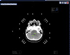
 (39Kbytes)
shows an axial CT of patient's head in a stereotactic frame. The white dots
outside the patient's head are part of the internal calibration of the stereotactic
frame.
Volumetric Stereotactic
Procedures
Volumetric stereotaxis is a
method for gathering, storing and reformatting imaging derived three dimensional
volumetric information defining an intracranial lesion with respect to the surgical
field. Most importantly, this information is displayed to the surgeon intraoperatively
and scaled to the actual size and location of the surgical field. With this technique
a surgeon can plan and simulate the surgical procedure beforehand, reach deep-seated
or centrally located brain tumors employing the safest and lest invasive route
possible.
Why is volumetric stereotaxis
necessary?
Intracranial mass lesions are
volumes in space. This is easily apparent on review of contiguous CT and MRI slice
images of the lesion. However, translation of this three dimensional information
from the imaging studies ( CT and MRI) to three dimensional surgical operating
space within the patient's head is difficult and imprecise during an open operation.
A surgeon may have difficulty in knowing where tumor ends and normal brain begins;
in spite of the fact that this information is usually clear on the imaging studies.
Indeed, there may even be difficulty in finding some subcortical tumors.
Without volumetric stereotaxis
three things are possible:
- A surgeon can get lost
attempting to find the tumor. Brain tissue is damaged unnecessarily. This
can result in neurologic deficit and prolonged and expensive rehabilitation
efforts.
- A surgeon can not tell
where tumor ends and normal brain tissue begins. Thus there is some risk that
the surgeon can resect normal brain tissue along with the tumor. In important
brain areas, this will also result in neurologic deficit.
- A surgeon performs a
subtotal removal of the lesion. Much tumor remains behind, will recur sooner
and require another operation or other treatments
- Complete removal of the
tumor.
What are the advantages
of volumetric stereotaxis?
Volumetric stereotaxis provides
the following major advantages to the surgeon in the management of intraaxial
brain lesions:
- It allows one to find
the lesion.
- It imparts a concept
of the three dimensional shape of the lesion which is to be removed.
- It allows preoperative
surgical simulation and surgical approach or trajectory planning with respect
to the configuration of the lesion and normal brain and vascular anatomy which
must be preserved. Thus the safest and most effective surgical approach may
be selected.
- It indicates by means
of a scaled real time display interactive software and stereotactic instrument
where tumor ends and normal brain begins.
Volumetric stereotaxis
has major advantages for the patient as well:
- The smallest possible
skin incision, craniotomy and brain incision. This minimizes injury to normal
brain tissue.
- Since the surgeon knows
exactly where tumor ends and normal brain begins, a more complete tumor removal
can be accomplished with much less risk to surrounding brain tissue.
- The postoperative neurologic
results are better than those associated with conventional (non-stereotactic,
non-volumetric) surgical techniques.
Volumetric stereotaxis
provides advantages to the insurance company since patients get out of the hospital
faster, do better neurologically, and back to work earlier. In a practical sense,
volumetric stereotaxis will save third party payors money because:
- Volumetric procedures
are less invasive than conventional intracranial neurosurgical procedures.
Post-operative results are better and patients get out of the intensive care
unit and out of the hospital faster. Less money is spent on ICU charges and
post-op hospital days. In a study done at the MAYO Clinic total hospital charges
including surgical fees for patients with astrocytic brain tumors undergoing
Computer-assisted stereotactic volumetric resection procedures were approximately
67% of the total hospital charges for conventional surgical procedures in
similar patients.
- Volumetric stereotactic
procedures require less time in the operating room ( 2-3 hours less in some
cases) than patients undergoing conventional neurosurgical procedures for
brain tumors. This is because the procedures are simulated on a computer system
beforehand and can proceed efficiently as planned. This saves money on operating
room charges.
- "Inoperable" tumors (inoperable
by conventional surgical techniques) can be resected with volumetric stereotactic
resection procedures. Frequently, these are deep seated-relatively benign
tumors in children and young adults. Many of these tumors can be cured with
volumetric stereotaxis.
- Neurologic results are
better, less patients require rehabilitation programs and return to work sooner.
Instrumentation required
for Volumetric stereotaxis:
- A capacious computer
and image processing system,
- Systems for data acquisition,
- Appropriate planning
and real-time display software,
- An interactive stereotactic
surgical system.
 (53.4Kbytes)
An view of treatment planning for volumetric stereotaxis.
Point-in-space and Volumetric
Stereotaxis
The difference between point-in-space
and volumetric stereotactic procedures. What Volumetric stereotactic procedures
provide that conventional point-in-space procedures do not.
Volumetric stereotactic
procedures are not to be confused with simple point-in-space stereotactic procedures,
which are employed for simple stereotactic biopsy, functional procedures, interstitional
irradiation of brain tumors or to correctly position a bone flap over a superficial
intracranial lesion or to find the superficial aspect of a deep-seated lesion.
Point-in-space procedures
are simple; volumetric procedures are mathematically complex and require a computer
system with specialized software to be done efficiently and safely.
Point in space procedures
can be done without a computer. However, computer-assisted surgical planning
with multimodality integration can be used in point-in-space stereotactic procedures
to make them more time efficient, accurate and safer. Volumetric procedures
cannot be performed without computer-assistance and intraoperative real-time
interaction.


 (39Kbytes)
shows an axial CT of patient's head in a stereotactic frame. The white dots
outside the patient's head are part of the internal calibration of the stereotactic
frame.
 (53.4Kbytes)
An view of treatment planning for volumetric stereotaxis.