 Ventricles of the Brain
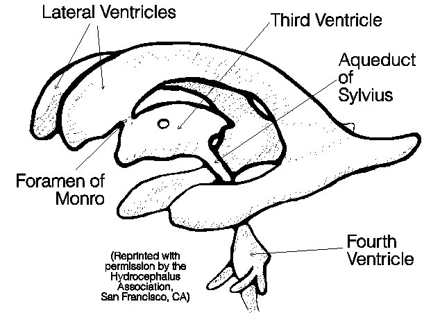
In children, brain tumors most commonly occur in the back of the brain (posterior fossa). As a tumor grows it may fill or compress the fourth ventricle, blocking the flow of cerebrospinal fluid. In other areas of the brain a tumor may similarly block or compress the ventricular system, causing hydrocephalus.
A shunt is a narrow, soft and pliable piece of tubing (approximately 0.25 cm. in diameter) which is surgically implanted into the ventricle through a small hole made in the skull.
All shunts have a valve system which regulates the pressure of the cerebrospinal fluid and prevents backward flow of fluid into the ventricles. Many shunts have reservoirs which can be used for removing CSF or administering drug therapy. A shunt may be pumped but this should only be done on a physician's order.
There are different types of shunts and placement procedures. To get a better idea of what a shunt looks like, ask to see one. Be sure to write down the name and type of the shunt your child has. This could be important information if your child runs into difficulty away from your medical centre.
![]() V-P Shunt
Ventricles of the Brain
V-P Shunt
Ventricles of the Brain
A Ventriculo-Peritoneal (VP) shunt is usually tried initially. Occasionally the abdomen cannot absorb fluid, and in these cases a Ventriculo-Atrial (VA) shunt system is used.
It may be useful for you to ask your neurosurgeon to show you an actual shunt. Be sure you know the name of your child's shunt.
Infants                 |      Toddlers          | Children and Adults
________________________|________________________|___________________________
Enlargement of the      | Head enlargement       | Vomiting
baby's head             |                        |
                        |                        |
Fontanelle is full and  | Vomiting               | Headache
tense when the infant   |                        |
is upright and quiet    |                        |
                        |                        |
Prominent scalp veins   | Headache               | Vision problems
                        |                        |
Swelling along the shunt| Irritability and/or    | Irritability and/or
tract                   | sleepiness             | tiredness
                        |                        |
Vomiting                | A loss of previous     | Personality change
                        | abilities (sensory or  |
                        | motor function)        |
                        |                        |
Irritability            | Seizures               | Seizures
                        |                        |
Sleepiness              |                        | Difficulty in waking
                        |                        | up or staying awake
                        |                        |
Downward deviation of   |                        | Loss of coordination
the eyes                |                        | or balance
                        |                        |
Seizures                |                        | Decline in academic
                        |                        | performance
                        |                        |
- - - Fever and redness along the shunt tract (with infection only) - - -
This list of symptoms is for your reference only, and is not a diagnostic aid. If
you are in doubt about your child's medical condition, consult your physician
immediately.This section on shunts was adapted in part from "About Hydrocephalus... A Book for Parents", a booklet produced by:
Hydrocephalus Association,
2040 Polk St. #342,
San Francisco, California
94109
(415)-776-4713
A unit or bag of blood drawn from a donor consists of almost 2 cups of whole blood and some anticoagulant and preservative. This blood can be given as it is, or it can be split into its component parts:
During surgery for brain tumors, some people require a blood transfusion. Transfusions may also be required if radiation or chemotherapy reduces the bone marrow's production of new blood.
Red cells carry oxygen from the lungs to the rest of the body, so they are very important. Lack of red blood cells causes anaemia. Your child may feel tired, fatigued or weak. These cells may need to be replaced with a packed red blood cell transfusion.
White blood cells are the major response by the immune system to infection. We are constantly exposed to bacteria or viruses which could make us quite ill but our immune system fights them off. If your child's white blood cell count is low, you may be asked to take special precautions to avoid infections such as avoiding other people with colds or infections, avoiding crowds (school, church, malls, movie theatres, etc.), and extra hand washing. At this time it is not possible to transfuse white blood cells. New drugs are being developed that can stimulate the body to make white blood cells. These growth factors (such as Neupogen [G-CSF, granulocyte colony stimulating factor]) must be given by injection and are usually only used if the white blood count is dangerously low after chemotherapy.
Likewise, blood platelets play an important part in protecting us. When we cut ourselves or bump a part of our body, the platelets collect at the site to help stop the bleeding. If their numbers are low, bleeding may occur which cannot be readily controlled. A blood transfusion may be necessary.
All transfusions of blood or blood products are given through an intravenous line (I.V or Port-a-Cath). During the transfusion, your child's pulse, blood pressure and temperature will be monitored closely to watch for any side effects to the blood being received. If a reaction does occur, he/she will be given a drug such as Tylenol or Benadryl (used with some allergic reactions) and the transfusion will be stopped. Fortunately, these reactions do not occur often.
Chemotherapy, often used for the treatment of brain tumors, may interfere with bone marrow function.
Once the treatment for the brain tumor has ended, the bone marrow's functional abilities gradually return to normal. During this time however, if your child is noted to be particularly pale, has less energy than is expected, bruises easily or experiences infection, you must promptly contact your oncologist. Blood or its by-products may yet be needed during this time as your child's bone marrow may not be able to produce enough on its own.
Across the country there are varying policies regarding the donation of blood for a family member. Should you have questions regarding policy and procedures, ask your physician and he/she will direct you to the appropriate source.
Being aware and sensitive to your child's visual abilities and/or restrictions will help you to determine if any change has occurred. Contact your doctor if you feel your child's vision has changed.
A change in vision is frequently a symptom that causes a person to seek medical help.
A person's visual system includes not only the eye itself but also the visual pathways that travel from the back of the eye (retina) all the way through to the back of the brain to the occipital lobes. Doctors who specialize in the eye and visual system are called Neuroopthalmologists.
The visual system is important in helping to make the initial diagnosis of a brain tumor and in continuing the management of the patient once the tumor has been treated.
Visual messages travel from the back of the eye along the two optic nerves and meet near the area of the pituitary gland called the optic chiasm. Here, the two nerves fuse together. Half of the pathways cross and the nerves travel back through the brain, through the temporal and parietal lobes, before arriving at the back of the brain (occipital lobes).
Visual fields are what the person can see in all directions with both eyes open and looking straight ahead. Each eye has a right and left visual field. These overlap the fields of the opposite eye.
When patients have difficulty behind the optic chiasm, the visual loss, because of the arrangement of the visual fibres, would be half or part of the vision on the one side so that the patient would have either the right side of the right eye vision lost and the right side of the left eye vision lost, or the reverse. See (Visual Fields, boxes C and D).
Tumors in the brain affecting the vision pathways behind the optic chiasm cause loss of the visual field on the opposite side in both eyes. For example, a tumor in the right side of the brain may cause loss of the left visual field in both eyes.
A tumor itself, or the pressure it causes, might hinder the nerve's ability to work, resulting in an imbalance in the action of the eye muscles. When this happens, one or both eyes may fall out of alignment with each other. Double vision may be the result.
The nerves involved are the third, fourth and sixth cranial nerves. If the third cranial nerve is involved, the eye may move outward and the eyelid may droop. The pupil of the eye may be large and not react well to light. If the fourth nerve is involved, the affected eye will move outward and up. They may not be able to look up. If the sixth nerve is involved, the eye will move inward toward the bridge of the nose.
Often, an intravenous contrast agent ("dye") must be injected in order to see the tumor more clearly as part of a CT or MRI scan.
It is very important that all patients lie absolutely still for these procedures. This can be difficult for the child under 5 years of age; thus, a mild sedative may be given to allow the child to sleep. The sedation used may be given by mouth (orally), injected into a muscle (intramuscularly) or injected into a vein (intravenously).
Some facilities have programs to prepare children to undergo these tests without sedation. Play therapy may be used. Please inquire at your hospital.
![]()
![]()
![]()