Gliomas
This is a name given to a family of brain tumors that are formed from the supporting cells of the nervous system. They do not arise from the neurons (nerves). These tumors account for over half of all brain tumors. Gliomas are subclassified into categories according to their cell type.
Gliomas may be given different names from centre to centre and over the past few years, patients have become increasingly confused when they read literature or speak to someone beyond their primary physician.
Generally, gliomas are classified according to whether they are slow or fast growing.
Astrocytomas
One third of all pediatric brain tumors are astrocytomas.
Grading Systems for Astrocytic Tumors
A number of different brain tumor classification systems have been proposed and continue to be used to group brain tumors into subtypes which help predict specific tumor growth characteristics.An accurate and reliable classification system used by physicians world-wide would be especially useful in the evaluation of different brain tumor treatments. The use of different classification systems for the same tumor has led to some confusion and it is recommended that you be aware of the type of tumor classification system used in the centre where your child is receiving treatment. The major classification systems used are outlined in the table below. The classification system used in this book is that of the World Health Organization's revised 1993 classification. Pilocytic astrocytoma is clearly differentiated from Astrocytoma (low grade) and the designation anaplastic astrocytoma and glioblastoma multiforme (GBM) are used to denote malignant astrocytic tumors. Please refer to the appendices for a complete schedule of classifications.
Comparison of a number of commonly used grading systems for astrocytomas
World Health            |               |
Organization            |  Kernohan     |    ..... St. Anne / Mayo Clinic .....
Designation             |  Grading      |    Designation  Histological Criteria
======================= |  ============ |    ===========--=====================
Pilocytic Astrocytoma   |               |    Astrocytoma  Grade 1 Zero criteria
                        |               |
Astrocytoma             |  Astrocytoma  |    Astrocytoma  One criteria usually
(low grade)             |  Grade 1      |    Grade 2      nuclear atypia
                        |  Astrocytoma  |
                        |  Grade 2      |
                        |               |
Anaplastic astrocytoma  |  Astrocytoma  |    Astrocytoma  Two criteria usually
                        |  Grade 3      |    Grade 3      nuclear atypia and
                        |               |                 mitotic activity
                        |               |
Glioblastoma Multiforme |  Astrocytoma  |    Astrocytoma  Three criteria usually
  (GBM)                 |  Grade 4      |    Grade 4      nuclear atypia,
                        |               |                 mitoses,
                        |               |                 endotherial proliferation
                        |               |                 and/or necrosis
Adapted from Kleihues et al Brain Pathology 3. 255-268 (1993)
Cerebellar Pilocytic Astrocytomas
These tumors are considered benign. First symptoms of these tumors are usually headache, vomiting or an unsteady gait. They occur equally between the sexes and at all ages in children, although they are unusual in infants. Approximately two-thirds of these tumors are cystic (have a fluid cyst associated with a solid mass) in nature.Management of the child with an astrocytoma usually involves also treating the problem of hydrocephalus. This is usually accomplished pre-operatively with steroids (dexamethasone, or the more common trade name of Decadron) which frequently relieve symptoms in 24-48 hours. A second approach to hydrocephalus infrequently requires the placement of a shunt in the days before surgery. See the chapter on shunts.
The goal of treatment of a cerebellar astrocytoma is complete surgical removal. This is achieved in a large majority of cases, and no further treatment is necessary. If total removal is not possible (for example it may have grown into the brain stem) radiation therapy may be necessary. Chemotherapy has not been found to be useful in the treatment of cerebellar astrocytomas. Pilocytic astrocytomas can occur in other parts of the brain and their treatment is usually surgery and radiation therapy if necessary.
Hemispheric Astrocytoma (low grade)
These are astrocytomas that are located in the hemispheres of the brain. They are most common in children between the ages of 8 and 12 years. First symptoms of these tumors usually include nausea, vomiting, headache and difficulties with vision.Surgical removal is the method of treatment for these astrocytomas. Radiation may also be necessary if the tumor is in an area of the brain responsible for such functions as speech, understanding or movement and it cannot be easily surgically removed.
Anaplastic Astrocytoma
The cells of these tumors are moderately fast growing and less well defined than an astrocytoma (low grade). Treatment involves removal of as much tumor as safely possible plus radiation and chemotherapy.Glioblastoma Multiforme (GBM)
These tumors may contain various cell types, hence the name "multiforme". These tumors are more common in the elderly.New data suggests that these tumors may be on the increase in the adult population. Surgery may be performed to remove as much tumor as possible. Radiation and chemotherapy are used to control growth of the tumor. Most of these tumors occur in the front portion of the brain (cerebral hemispheres).
The cells of these tumors grow quickly, are not well defined, and they may frequently spread throughout the brain.
Brain Stem Gliomas
Brain stem gliomas are astrocytomas that usually start in the pons. Ten to twenty-five percent (10%-25%) of pediatric brain tumors will be brain stem gliomas. These tumors occur equally between sexes and most often occur between the ages of 5-10 years. Brain stem gliomas are located in an area of the brain and spinal cord that is responsible for many vital body functions. These include vision, balance, strength, gagging, coughing and swallowing.Because of the location of these tumors, surgery is rarely an option (except for biopsy and draining of cyst). Radiation to the mass is the usual method of treatment. Some centres may add chemotherapy before or after radiation treatment.
Optic Nerve Gliomas
Optic nerve gliomas represent four to six percent (46%) of all pediatric brain tumors. They are located along the optic nerves, the optic chiasm and the hypothalamus. See Eye Signs and Symptoms in Brain Tumors.These tumors are usually slow growing (astrocytomas). Symptoms will vary depending on location but may include decreased vision, double vision and papilledema (swelling of the optic nerve). If vision has been lost, surgery may be considered. If visual function remains, there will be careful follow-up with imaging studies done at regular intervals. Radiation therapy may be an option. For tumors that involve the hypothalamus and the third ventricle, there may be signs of hydrocephalus as well as hormone imbalance. Surgical treatment of hydrocephalus may be considered. Some tumors located here respond well to chemotherapy and it may be considered.
Oligodendrogliomas
These tumors are not common in children and are slow growing. The mass usually appears calcified (calcium deposits) on imaging (CT scan or MRI scan) showing that it has been there for many years. These tumors arise from the oligodendroglial cells which make up the myelin that insulates nerve fibres. Symptoms of these tumors may include seizures, headache and vision problems. Treatment usually involves surgical removal along with a course of radiation and/or chemotherapy if sub-totally removed.Ependymomas
These tumors arise from the cells lining the ventricles (hollow channels) of the brain (most commonly the fourth ventricle). As these tumors grow and fill the ventricle, they obstruct the flow of cerebrospinal fluid (CSF) through the brain. These tumors appear more commonly among younger children. Symptoms of this tumor include headache, nausea, vomiting, balance problems and visual disturbances.Hydrocephalus, caused by the blocking of the flow of cerebrospinal fluid, may make shunting necessary. Total surgical removal is not always possible, and treatment often involves radiation therapy and/or chemotherapy.
Gangliogliomas
Gangliogliomas are generally slow growing tumors and may occur anywhere in the brain although the temporal lobe is the most common site. The average age at the time of diagnosis is 12 years. Many children are diagnosed after a long history of seizures that have been difficult to treat. Intellectual and behavioral difficulties may be present. Treatment is usually total removal by surgery.Mixed Gliomas
You may hear this term used. A tumor may be composed of two or more cell types (as seen under the microscope). The tumor is generally named for the type of glioma cells which grow the fastest.
Primitive Neuroectodermal Tumors (PNET)
Medulloblastoma
Twenty percent (20%) of all pediatric brain tumors are PNETs. They are most commonly found in the cerebellum and are called medulloblastomas when they occur here. They are most commonly found in the cerebellum. These tumors are twice as common in boys, with diagnosis frequently between the ages of 4 and 8. These tumors are fast growing and symptoms include a short, progressive history of headaches, vomiting, loss of appetite and coordination difficulties. Spread outside of the brain and spinal cord is rare although it may occur. Very often, these tumors have spread within the central nervous system before diagnosis. Spread through the CSF is common (carcinomatous meningitis). The extent of the tumor is often classified according to a staging system. This system is outlined below and is an important consideration when planning appropriate treatment:
Stage | Description
======  =================================================================
T1      Tumor is less than 3cm. in diameter and does not go beyond the
        midline of the brain, the roof of the fourth ventricle or the
        cerebellar hemispheres.

T2      Tumor is greater than 3 cm. in diameter and grows into neighboring
        brain or spinal cord, or partly fills the fourth ventricle.

T3a     Tumor grows into two neighboring parts of the brain or spinal cord
        or completely fills in the fourth ventricle producing signs of
        hydrocephalus.

T3b     Tumor grows from the floor of the fourth ventricle or brain stem
        and fills the fourth ventricle, producing signs of hydrocephalus.

T4      Tumor spreads to involve the third ventricle or midbrain or upper
        cervical cord.

M0      No evidence of spread around the brain or to any other part of the
        body

M1      Microscopic tumor cells found in cerebrospinal fluid.

M2      Tumor seeding (separate from primary tumor) found in cerebellum,
        subarachnoid space or in the third or lateral (on either side)
        ventricles.

M3      Tumor seeding (separate from primary tumor) found in spinal
        subarachnoid space.

M4      Tumor spread outside the cerebrospinal system.
Chang CH, Housepian EM, Herbert C Jr.: An operative staging system and a megavoltage radiotherapeutic technique for cerebellar medulloblastomas, Radiology 93: 1351-1359, 1969
It is important to be aware of the above stages as they may be discussed with you in terms of treating your child's tumor.Treatment almost always involves surgery with the goal of removing all (gross total) or as much as possible (subtotal) of the tumor. Radiation is very effective against this tumor and is generally done to the entire head and spinal cord because of the high possibility of seeding malignant cells by way of the cerebrospinal fluid. Chemotherapy has been shown to be effective. After your child has had his/her tumor staged, the treatment best fitted for his/her tumor will be used. An effective chemotherapy protocol has been developed and although we do not yet know the long-term results of its use, the outlook for this type of tumor has improved dramatically over the last ten years. Shunting may be necessary to treat hydrocephalus caused by the tumor interrupting the pathways of the cerebrospinal fluid.
Other Primitive Neuroectodermal Tumors
- cerebral neuroblastoma (located in hemispheres)
- ependymoblastoma (arises from ventricles)
- pineoblastoma (occurs in area of pineal gland)
Pineal Region Tumors
The pineal gland is located centrally in the brain and is an outpouching of the third ventricle. Hydrocephalus is usually a presenting symptom as the cerebrospinal fluid pathway may be blocked. The optic (vision) pathways may also be involved and there may be some double vision. Surgery is sometimes possible or a biopsy may be performed to obtain a diagnosis. Some of these tumors are sensitive to radiation. A shunt may be necessary to treat the hydrocephalus, although hydrocephalus may be initially controlled by the use of steroids. Some germ cell tumors may be treated with chemotherapy.There are four categories of pineal region tumors:
- Germ cell tumors:
- fast growing:
- choriocarcinoma
- embryonal carcinoma
- endodermal sinus tumor
- not as fast growing:
- germinomas
- slow growing:
- dermoid cysts
- epidermoid cysts
- teratomas
Dermoid and Epidermoid Cysts
These cysts develop from congenital tissue (formed before birth). Epidermoid cysts contain keratin, cellular debris and cholesterol. Dermoid cysts contain hair and sebaceous (sweat) glands. These masses occur in central areas of the brain such as the hypothalamic region, the vermis of the cerebellum and the pineal region. The bones of the skull (not involving the brain itself) and the spine may also be involved. These cysts are treated with surgery and complete removal is usually possible.Teratomas
This is a congenital tumor which is made up of elements from the three primary cell layers: Ectoderm (skin and nervous system), Mesoderm (muscle, bone and cartilage) and Endoderm (gut lining). These are the earliest cell types in development. It is most commonly a tumor of the pineal gland or lower spinal cord in the central nervous system, but may be found near the base of the brain near the pituitary gland, or the base of the third ventricle. Treatment is usually surgery.
- fast growing:
- Pineal cell tumors:
- fast growing: pinealoblastomas
- slow growing: Pinealocytomas
These tumors arise from the actual pineal cells of the pineal gland itself. If the cells are dividing quickly, they are considered pinealoblastomas, whereas those tumors with slow growing cells are called pinealomas.
Treatment is usually biopsy, or if small, surgical removal. Radiation therapy follows and some centres are using chemotherapy.
- Glial cell tumors:
Any glial tumor, both slow and fast growing, can be found in the pineal
region. These are discussed earlier in this chapter (e.g. astrocytomas).
- Miscellaneous tumors:
These tumors are almost all slow growing:
- cysts: accumulation of abnormal fluid enclosed in a lining.
- meningiomas: a tumor with distinct borders usually causing symptoms by pressure on the neighboring area of the brain (discussed later in this chapter).
Craniopharyngiomas
These slow growing tumors account for approximately nine percent (9%) of all pediatric brain tumors and usually occur in children between 5 and 10 years of age. These tumors arise from cells along the pituitary stalk and may grow up and involve the hypothalamus, optic nerve pathways and the third ventricle. These tumors are cystic in nature and slow growing. At diagnosis, children may have signs of increased pressure in the brain (third ventricle blocked), visual loss (either decreased vision or visual field problems) and hormone difficulties (growth delay, thyroid deficiency, sexual delay). Complete removal of this tumor is possible if it is in a favorable location. Otherwise, radiation therapy may be used. All of these children will require long term follow-up by an endocrinologist (a physician who studies the hormone systems of the body) and a neuroopthalmologist. Some of these children will require neuropsychological follow-up. Usually, lifelong hormone replacement is necessary.
Choroid Plexus Papillomas
The choroid plexus is an infolding of the lining of the ventricular surface. This surface has many blood vessels. The choroid plexus is involved in the production of most of the cerebrospinal fluid. A child with a choroid plexus papilloma usually has marked hydrocephalus due to obstruction of flow and sometimes an overproduction of cerebrospinal fluid.The most common location for this tumor is in the lateral ventricles (the ventricles on either side of the brain). These tumors account for one to three percent (1%-3%) of pediatric brain tumors and are most common in children under the age of 2 years. They make up eight percent (8%) of brain tumors diagnosed in newborn babies.
These tumors are separated into 2 categories as follows:
- Papillomas: slow growing (vast majority)
- Carcinomas: fast growing (rare)
Treatment for these tumors involves complete surgical resection (removal) for papillomas, whereas carcinomas require surgery, radiation and chemotherapy. On rare occasions, carcinomas may spread to other areas.
Meningiomas
This is a rare tumor of childhood. It is usually a benign tumor that grows within the skull but on the outside of the brain. On rare occasions it may grow within the ventricles. Surgical removal may be curative. Radiation may be used if complete removal is impossible, or the tumor is fast growing. Meningiomas are more common in adults, especially in old age.
Colloid Cysts
These cysts usually occur in the third ventricle and can cause hydrocephalus. They contain embryonic tissue (tissue formed before birth). Treatment includes surgery and sometimes shunting.
Chordomas
These tumors develop from remnants of embryonic tissue. They are slow growing and usually do not produce symptoms until middle age. These tumors can arise in the skull bones at the base of the brain, or in the vertebral bones in the neck or sacrum. Treatment is surgery and radiation therapy.
Lymphomas
Lymphomas arise from specialized cells of the lymphatic system and occur both in the brain and spinal cord. The incidence is increased in patients after organ transplantation and patients whose immune system is depressed (e.g. AIDS). These tumors are frequently responsive to steroid medication. Surgery and radiation may be indicated along with chemotherapy.Primary brain lymphomas are rare in children, but lymphomas starting elsewhere in the body may spread to the brain or spinal fluid.
Pituitary Adenomas
These are tumors of the pituitary gland and can be divided into two groups. The first group shows signs of a space-occupying lesion where the other shows signs of abnormal activity of the gland.
- Space-occupying lesions (nonfunctioning tumor) - Symptoms of these
tumors result from pressure on neighboring structures. They may produce
visual disturbances if they compress the visual pathways. Hormone
secretion by the pituitary gland can be decreased by compression caused by
the growing tumor. This can result in signs of hypopituitarism such as a lack
of menstrual periods, reduction of body hair, increased sensitivity to the
cold and decreased function of glands stimulated by the pituitary. Treatment
is surgical removal and radiation is sometimes used, either alone or in
conjunction with surgery.
- Secreting Pituitary Tumors (functioning tumors) - Pituitary tumors that
contain hormone-secreting cells often produce clinical symptoms before
tests can actually show enlargement of the pituitary gland.
- Prolactin Secreting Adenomas - These tumors alter hormones resulting in
lack of menstrual periods and infertility in women and impotence in men.
Galactorrhea (milk coming from the breasts) can also occur.
- Growth Hormone Secreting Adenomas - This tumor secretes excessive
amounts of growth hormone. In children, giantism results and in adults,
acromegaly results. Common features of giantism include rapid growth,
joint and muscle tenderness, exercise intolerance and fatigue. In the adult,
changes can occur slowly over years. Other conditions that may occur
include diabetes mellitus, kidney problems, increased metabolic rate and
hypertension (high blood pressure).
- In young children, hypopituitary function (lack of) can result in decreased
overall growth and the child doesn't grow at a normal rate.
- ACTH (Adrenocorticotropic) Secreting Adenomas - These tumors produce
ACTH which stimulates the production of cortisol from the adrenal gland.
This overproduction of cortisol impairs the body's response to injury and
infection. It can deplete the body's potassium while retaining sodium and
water. This tumor may result in a change in the body's fat distribution
(Cushingoid appearance).
Familial Diseases Associated with Brain Tumors
Genetic counselling is advised in all of the following conditions. Ask for a referral if one is not offered.
Neurofibromatosis
Sometimes called Von Recklinghausen's Disease.- Type 1 - This is a genetically dominant disorder that is passed from one
generation to the next. Neurofibromas are slow growing tumors located on
or beneath the skin. They may also occur in deeper layers as well as in
internal organs. Neurofibromas are composed of tissues from the nervous
system, (neuro) and fibrous tissue(fibroma), which form the support
structure of the nerve. The skin, bones, endocrine glands and nervous
system are sites of congenital abnormality.
Many types of brain tumors can be associated with neurofibromatosis. Most commonly, these are optic nerve tumors, gliomas in other areas, meningiomas and neurofibromas, especially along the spinal cord. These tumors are usually benign. Surgery and/or radiation therapy is sometimes indicated, when the tumor occurs within the central nervous system (brain and spinal cord).
- Type 2 - This is different from Type 1 but is also a genetically dominant disorder. It is usually diagnosed by the presence of acoustic neuromas (Schwannomas) on the vestibulo-cochlear nerves on one or both sides of the brain. It usually presents as deafness. Other gliomas may be present. Surgery is often indicated.
The Neurofibromatosis Society of Ontario,
923 Annes St.,
Whitby, Ontario L1N 5K7
(The society serves all of Canada)or
Neurofibromatosis, Inc.
8855 Annapolis Rd., Suite 110,
Lanham, MD 20706-2924
U.S.A.- Type 1 - This is a genetically dominant disorder that is passed from one
generation to the next. Neurofibromas are slow growing tumors located on
or beneath the skin. They may also occur in deeper layers as well as in
internal organs. Neurofibromas are composed of tissues from the nervous
system, (neuro) and fibrous tissue(fibroma), which form the support
structure of the nerve. The skin, bones, endocrine glands and nervous
system are sites of congenital abnormality.
Hippel-Lindau Disease
This is a genetically dominant disorder that may not be evident until the patient has children of his/her own. Hemangioblastomas, often in the cerebellum, are the most common brain tumors associated with this disease. Hemangioblastomas are seen associated with the retina of the eye and, if not treated, can lead to blindness. Treatment is usually surgery for the brain tumor. Laser coagulation can be used for retinal lesions.Tuberous Sclerosis
This is a genetically dominant disorder which usually starts with seizures. Tumors associated with it are usually giant cell astrocytomas in the region of the third ventricle, presenting with hydrocephalus. It can cause tumors in organs other than the brain. Treatment includes control of seizures and possibly surgery.
Tuberous Sclerosis Association of Canada,
2443 New Wood Dr.
Oakville, Ontario
L6H 5Y3or
National Tuberous Sclerosis Foundation
8000 Corporate Dr.,
Suite 120,
Landover, MD 20785
U.S.A.
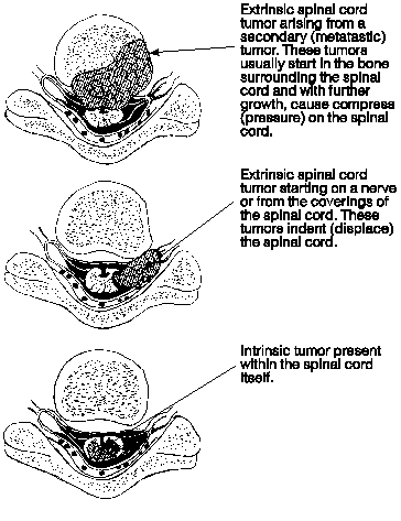
Spinal Cord Tumors
Tumors which occur in the brain can also occur in the spinal cord, the most common ones being in the glioma family (astrocytomas, ependymomas).
- Intrinsic - These occur in the substance of the spinal cord and may be invasive. Treatment is surgery and/or radiation.
- Extrinsic - These occur outside the spinal cord and exert pressure on the
cord as they grow. The symptoms of these tumors are related to pressure.
These tumors can arise from:
- dura (meningiomas) or nerves (neurofibromas and Schwannomas) surrounding the spinal cord. These tumors indent the spinal cord like a marble.
- metastatic deposits from tumors elsewhere in the body. These tumors start in the bone surrounding the spinal cord and may compress the spinal cord from a number of sides.
Treatment is surgery and/or radiation.
 Spinal Cord and Coverings